James Priest couldn’t make sense of it. He was examining the DNA of a desperately ill baby, searching for a genetic mutation that threatened to stop her heart. But the results looked as if they had come from two different infants.
“I was just flabbergasted,” said Dr. Priest, a pediatric cardiologist at Stanford University.
The baby, it turned out, carried a mixture of genetically distinct cells, a condition known as mosaicism. Some of her cells carried the deadly mutation, but others did not. They could have belonged to a healthy child.
We’re accustomed to thinking of our cells sharing an identical set of genes, faithfully copied ever since we were mere fertilized eggs. When we talk about our genome — all the DNA in our cells — we speak in the singular.
But over the course of decades, it has become clear that the genome doesn’t just vary from person to person. It also varies from cell to cell. The condition is not uncommon: We are all mosaics.
For some people, that can mean developing a serious disorder like a heart condition. But mosaicism also means that even healthy people are more different from one another than scientists had imagined.
Magical Mystery
In medieval Europe, travelers making their way through forests sometimes encountered a terrifying tree.
A growth sprouting from the trunk looked as if it belonged to a different plant altogether. It formed a dense bundle of twigs, the sort that people might fashion into a broom.
Germans call it Hexenbesen: witches’ broom. As legend had it, witches used magic spells to conjure the brooms to fly across the night sky. The witches used some as nests, too, leaving them for hobgoblins to sleep in.
In the 19th century, plant breeders found that if they cut witches’ broom from one tree and grafted it to another, the broom would grow and produce seeds. Those seeds would sprout into witches’ broom as well.
Today you can see examples of witches’ broom on ordinary suburban lawns. Dwarf Alberta spruce is a landscaping favorite, growing up to ten feet high. It comes from northern Canada, where botanists in 1903 discovered the first known dwarf clinging to a white spruce — a species that can grow ten stories tall.
Pink grapefruits arose in much the same way. A Florida farmer noticed an odd branch on a Walters grapefruit tree. These normally bear white fruit, but this branch was weighed down with grapefruits that had pink flesh. Those seeds have produced pink grapefruit trees ever since.
Charles Darwin was fascinated by such oddities. He marveled at reports of “bud sports,” strange, atypical blooms on flowering plants. Darwin thought they held clues to the mysteries of heredity.
The cells of plants and animals, he reasoned, must contain “particles” that determined their color, shape and other traits. When they divided, the new cells must inherit those particles.
Something must scramble that heritable material when bud sports arose, Darwin declared, like “the spark which ignites a mass of combustible matter.”
Only in the 20th century did it become clear that this combustible matter was DNA. After one cell mutates, scientists found, all its descendants inherit that mutation.
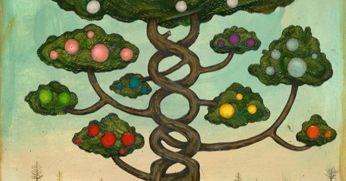
Witches’ broom and bud sports eventually came to be known as mosaics, after the artworks made up of tiny tiles. Nature creates its mosaics from cells instead of tiles, in a rainbow of different genetic profiles.
Before DNA sequencing was commonplace, scientists struggled to tell the genetic differences between human cells. Cancer offered the first clear evidence that humans, like plants, could become mosaics.
In the late 1800s, biologists studying cancer cells noticed that many of them had oddly shaped chromosomes. A German researcher, Theodor Boveri, speculated at the turn of the century that gaining abnormal chromosomes could actually make a cell cancerous.
As soon as Boveri floated his theory, he faced intense opposition. “The skepticism with which my ideas were met when I discussed them with investigators who act as judges in this area induced me to abandon the project,” he later said.
Boveri died in 1915, and it took nearly five decades for scientists to discover he was right.
David A. Hungerford and Peter Nowell found that people with a form of cancer called chronic myelogenous leukemia were missing a substantial chunk of chromosome 22. It turned out a mutation had moved that chunk over to chromosome 9. The cells that inherited that mutation became cancerous.
It’s hard to think that a tumor might have anything in common with a pink grapefruit. Yet they are both products of the same process: lineages of cells that gain new mutations not found in the rest of the body.
Some skin diseases proved to be caused by mosaicism, too. Certain genetic mutations cause one side of the body to become entirely dark. Other mutations draw streaks across the skin.
The difference is in the timing. If a cell gains a mutation very early in development, it will produce many daughter cells that will end up spreading across much of the body. Late-arising mutations will have a more limited legacy.
A Brain Biography
Dr. Walsh and his colleagues have found evidence of mosaicism in some very unexpected places.
They investigated a mysterious disorder called hemimegalencephaly, which causes one side of the brain to become overgrown. The researchers examined tissue from patients who had brain surgery to treat the seizures triggered by hemimegalencephaly.
Some of the brain cells in the patients — but not all of the cells — shared the same mutant genes. It’s possible that these mutant neurons multiplied faster than others in the brain, triggering one side to become enlarged.
Preliminary studies suggest that mosaicism underlies many other diseases. Last year, Christopher Walsh, a geneticist at Harvard University, and his colleagues published evidence that mosaic mutations may raise the risk of autism.
But scientists are also finding that mosaicism does not automatically equal disease. In fact, it’s the norm.
When a fertilized egg — known as a zygote — starts dividing in the womb, many of its early descendant cells end up with the wrong number of chromosomes. Some are accidentally duplicated, and others lost.
Most of these unbalanced cells divide only slowly or die off altogether, while the normal cells multiply far faster. But a surprising number of embryos survive with some variety in their chromosomes.
Markus Grompe, a biologist at Oregon Health & Science University, and his colleagues looked at liver cells from children and adults without liver disease. Between a quarter and a half of the cells were aneuploids, typically missing one copy of one chromosome.
Along with altered chromosomes, human embryos also gain smaller mutations in the genome. Stretches of DNA may be copied or deleted. Single genetic letters may get incorrectly reproduced.
It wasn’t possible to study such molecular changes accurately until DNA-sequencing technology became sophisticated enough.
In 2017, researchers at the Wellcome Trust Sanger Institute in England examined 241 women, sequencing batches of white blood cells from each. Every woman had acquired about 160 new mutations, each present in a sizable fraction of her cells.
The women gained these mutations as embryos, the scientists theorized, with two or three new mutations arising each time a cell divided. As those new mutations occurred, the embryonic cells passed them all down to their descendants, a mosaic legacy.
Dr. Walsh and his colleagues have discovered intricate mosaics in the brains of healthy people. In one study, they plucked neurons from the brain of a 17-year-old boy who had died in a car accident. They sequenced the DNA in each neuron and compared it to the DNA in cells from the boy’s liver, heart and lungs.
Every neuron, the researchers found, had hundreds of mutations not found in the other organs. But many of the mutations were shared only by some of the other neurons.
It occurred to Dr. Walsh that he could use the mutations to reconstruct the cell lineages — to learn how they had originated. The researchers used the patterns to draw a sort of genealogy, linking each neuron first to its close cousins and then its more distant relatives.
When they had finished, the scientists found that the cells belonged to five main lineages. The cells in each lineage all inherited the same distinctive mosaic signature.
Even stranger, the scientists found cells in the boy’s heart with the same signature of mutations found in some brain neurons. Other lineages included cells from other organs.
Based on these results, the researchers pieced together a biography of the boy’s brain.
When he was just an embryonic ball in the womb, five lineages of cells had emerged, each with a distinct set of mutations. Cells from those lineages migrated in different directions, eventually helping to produce different organs — including the brain.
The cells that became the brain turned into neurons, but they did not all belong to the same family. Different lineages merged together. In essence, the boy’s brain was made of millions of mosaic clusters, each composed of tiny cellular cousins.
It’s hard to say what these mosaic neurons mean to our lives — what it means for each of us to have witches’ broom growing in our skulls. “We don’t know yet whether they have any effect on shaping our abilities or challenges,” said Dr. Walsh.
What we do know is that mosaicism introduces randomness into the development of our brains. Mutations, which arise at random, will form different patterns in different people. “The same zygote would never develop exactly the same way twice,” said Dr. Walsh.
A Heart in Pieces
As ubiquitous as mosaicism may be, it’s still easy to overlook — and surprisingly hard to document.
Astrea Li, the infant examined by Dr. Priest at Stanford, had gone into cardiac arrest the day she was born. Her doctors put a defibrillator in her heart to shock it back into the proper rhythm.
Dr. Priest sequenced Astrea’s genome to search for the cause of her disorder. He concluded that she had a mutation in one copy of a gene called SCN5A. That mutation could have caused her trouble, because it encodes a protein that helps trigger heartbeats.
But when Dr. Priest ran a different test, he couldn’t find the mutation.
To get to the bottom of this mystery, he teamed up with Steven Quake, a Stanford biologist who had pioneered methods for sequencing the genomes of individual cells. Dr. Priest plucked 36 white blood cells from the child’s blood, and the scientists sequenced the entire genome of each cell.
In 33 of the cells, both copies of a gene called SCN5A were normal. But in the other three cells, the researchers found a mutation on one copy of the gene. Astrea had mosaic blood.
Her saliva and urine also turned out to contain mosaic cells, some of which carried the mutation. These findings demonstrated that Astrea had become a mosaic very early in her development.
The skin cells in her saliva, the bladder cells in her urine and her blood cells each originated from a different layer of cells in two-week-old embryos.
Astrea’s SCN5A mutation must have originated in a cell that existed before that stage. Its daughter cells later ended up in those three layers, and ultimately in tissues scattered throughout her body.
They might very well have ended up in her heart, too. And there the mutation could have theoretically caused Astrea’s problems.
While Dr. Priest was reconstructing Astrea’s mosaic origins, she was recovering from the surgery to implant her defibrillator. Her parents, Edison Li and Sici Tsoi, brought her home. And for a few months, it seemed she was out of the woods.
One day, however, her defibrillator sensed an irregular heartbeat and released a shock — along with a wireless message to Astrea’s doctors.
Back at the hospital, doctors discovered a new problem: her heart had become dangerously enlarged. Researchers have linked mutations in the SCN5A gene to the condition.
Her heart soon stopped. Her doctors attached a mechanical pump, and soon a donated heart became available.
Astrea underwent transplantation surgery and recovered well enough to go home. She went on to enjoy a normal childhood, performing cartwheels with her sister and listening obsessively to the soundtrack of “Frozen.”
The transplant did not just give Astrea a new lease on life. It also gave Dr. Priest a very rare chance to look at a mosaic heart up close.
The transplant surgeons had clipped some pieces of Astrea’s cardiac muscle. Dr. Priest and his colleagues extracted the SCN5A gene from the cells taken from different parts of her heart.
On the right side of the heart, he and his colleagues found that more than 5 percent of the cells had mutant genes. On the left, nearly 12 percent did.
To study the effect of this mosaicism, Dr. Priest and his colleagues built a computer simulation of Astrea’s heart. They programmed it with grains of mutant cells and let it beat.
The simulated heart thumped irregularly, in much the same way Astrea’s had.
The experience left Dr. Priest wondering how many more people might be at risk from a hidden mix of mutations.
Unless he winds up with another patient like Astrea, we may never find out.
An earlier version of this article misstated the year in which Theodor Boveri died. It was 1915, not 1914.
Source : Nytimes


